A Hidden Organ Scientists Missed for Centuries Has Finally Been Found
Last updated on
For generations, the human body has been presented as one of science’s most complete achievements. Medical textbooks outline organs with precision. Diagrams appear final and authoritative. Students are taught that while treatments and technologies evolve, the basic map of the body has long been settled. That belief makes what happened in 2020 so startling. In the midst of routine cancer research, scientists unintentionally uncovered something no one was actively searching for. Hidden deep within the human head was a structure large enough to influence daily functions like swallowing, speaking, and breathing. It had existed in every patient examined, yet it had gone unrecognized for centuries. Researchers had not only found a new anatomical structure, but one that could meaningfully change how cancer patients are treated. The discovery was not dramatic or theatrical. There was no single eureka moment. Instead, it unfolded quietly, through repeated observations, careful verification, and a growing realization that something fundamental had been overlooked. What followed challenged assumptions about modern medicine and reminded scientists and the public alike that even in the twenty first century, the human body still has secrets.An Unexpected Finding During Cancer Research
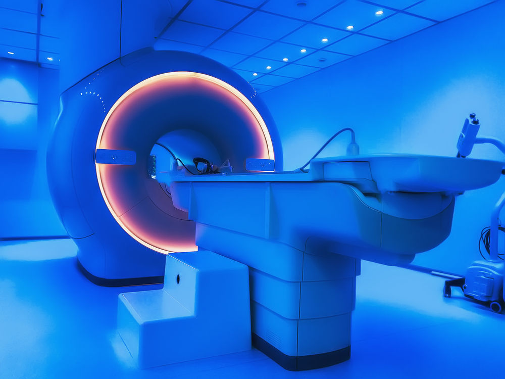
The discovery began at the Netherlands Cancer Institute, where researchers were studying prostate cancer patients using advanced imaging technology. Their focus was narrow and practical. They wanted to detect tumors with greater accuracy and understand how cancer spreads throughout the body. The scans they used were designed to highlight cancer cells by making them glow. Yet as clinicians reviewed images from patient after patient, something unusual began to appear. Two symmetrical areas in the head consistently lit up on the scans. These glowing regions were not random. They appeared in the same place every time, deep behind the nose. Initially, the findings were easy to dismiss. Medical imaging often produces artifacts, shadows, or anomalies. A single unexpected image might be ignored. Even a few could be written off as coincidence. But as the pattern repeated itself across dozens of patients, the explanation became harder to ignore. Eventually, nearly one hundred individuals displayed the same glowing structures in the same location. At that point, the research team began to suspect they were not looking at a technical glitch or a rare anatomical variation. They were looking at something real.The Imaging Technology That Made It Visible
Proving It Was Not an Illusion

Why Anatomy Missed This for So Long

Understanding the Role of Salivary Glands

The Connection to Radiation Therapy Side Effects

A Discovery With Immediate Real World Impact
Many scientific discoveries take decades to influence everyday medical practice. This one is different. Because radiation therapy is already carefully mapped and personalized for each patient, incorporating protection for the tubarial glands could happen relatively quickly. The knowledge does not require new equipment or experimental treatments. It requires awareness. For patients, the potential benefits are profound. Reduced dryness, improved swallowing, and better speech function can dramatically change recovery and long term wellbeing. Quality of life is often as important as survival, especially for patients who live many years after cancer treatment. This makes the discovery of the tubarial glands more than an academic curiosity. It represents a practical step forward in patient care.
Are the Tubarial Glands Truly a New Organ
Not all experts agree on how to classify these structures. Some anatomists caution against labeling them as an entirely new organ system. The human body contains hundreds, possibly thousands, of minor salivary glands scattered throughout the mouth and throat. From this perspective, the tubarial glands may represent an unusually organized cluster rather than a completely new category. Others point out that the original study population consisted largely of male patients, due to the prostate cancer focus. Broader studies involving women, different age groups, and diverse populations will be necessary to fully understand the universality of these glands. This skepticism reflects the careful process of scientific validation. Debate and replication are essential before new ideas become established knowledge. Even so, most experts agree on one thing. Whether classified as a new organ or a newly recognized glandular system, the tubarial glands are clinically significant and deserve attention.Lessons About Scientific Certainty
Beyond its medical implications, the discovery carries a broader lesson about science itself. Throughout history, there have been moments when entire fields believed they were complete. Physics at the end of the nineteenth century famously assumed there was little left to discover. Neuroscience once viewed brain function as rigid and localized. In each case, new tools and perspectives revealed deeper complexity. Anatomy has long been treated as settled science. The idea that a structure large enough to influence speech and swallowing could remain hidden until recently challenges that assumption. This does not indicate failure. It demonstrates how knowledge evolves alongside technology and curiosity.
The Symbolic Weight of Hidden Anatomy
There is also something quietly symbolic about where these glands are located. Positioned behind the nose and near the throat, they sit at the intersection of breath, voice, and sensation. This region plays a central role in communication and perception, yet it operates largely without conscious awareness. Science does not assign symbolic meaning to anatomy. Still, it is difficult to ignore the resonance of discovering something essential that has been quietly supporting human function all along. The tubarial glands remind us that importance does not always announce itself. Some of the most critical systems operate silently, unnoticed until something goes wrong.New Directions for Future Research
The Human Body as an Unfinished Map
The discovery of the tubarial salivary glands serves as a reminder that the human body is not a static diagram. It is a living system, shaped by evolution and revealed gradually through observation and inquiry. Accidental discoveries have always played a role in scientific progress. Penicillin, X rays, and anesthesia all emerged from unexpected observations. The tubarial glands now join that lineage. They show how paying attention to anomalies, rather than dismissing them, can lead to meaningful breakthroughs.Some of the links I post on this site are affiliate links. If you go through them to make a purchase, I will earn a small commission (at no additional cost to you). However, note that I’m recommending these products because of their quality and that I have good experience using them, not because of the commission to be made.


 JOIN OVER
Comments